The calf gets the attention. It is not the whole story
What muscles are weak in clubfoot (club foot, talipes) is a more specific question than the calf. Everyone notices the smaller calf, because it is visible and because someone points it out in a changing room. Underneath it there is a more specific pattern of weakness, and knowing which muscles are involved changes what is worth training and what is a waste of six months. Four groups, opened in the order they matter.
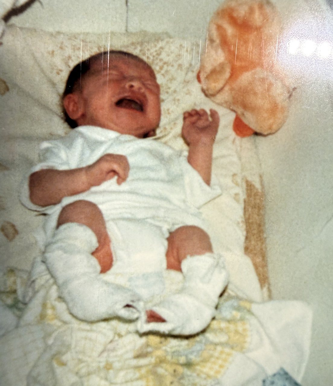

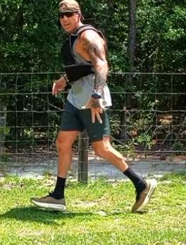
Four muscle groups. Open the ones relevant to you. Almost everyone has the first, and the second is the one most often missed entirely.
The calf (gastrocnemius and soleus) Universally affected, partly trainable, never symmetrical. Group 12 min
The calf on an affected side is smaller in essentially every treated clubfoot, and the difference persists for life regardless of training. Measured differences of one to three centimeters in circumference are typical. It is not disuse. The muscle was built smaller, and it was built smaller before anybody touched it.1
Imaging in Ponseti-treated children found the plantarflexor muscles smaller on the affected side and the Achilles tendon larger, with the size of the difference showing no relationship to age.2 Less muscle, more tendon, and the proportion set early.
What this costs is push-off. The calf drives the final propulsive phase of every step, so a smaller one means less power from that side, every stride, forever. On a walk to the shops that is invisible. Over a marathon, or a duty day in boots, it is the reason one leg tires first.
Worth training anyway. Strength and endurance respond well even where size does not. See what training actually reaches.
The peroneals (the outside of the shin) The group nobody mentions, and the one that explains rolled ankles. Group 22 min
The peroneal muscles run down the outside of the lower leg and turn the foot outward, against the tibialis anterior and posterior pulling it in. That balance is the whole story here, and when it tips the clinical name for it is dynamic supination: the foot rolls inward as it swings through, most visibly in a child learning to walk.3
What nobody has published, as far as I can find, is a study measuring evertor strength in treated adult clubfoot. The imbalance is well documented and this specific muscle group is not, so treat what follows as mechanism and experience instead of measurement.
The consequence is a foot that resists turning outward and has less active defense against rolling inward. If you roll your ankle more than seems reasonable, or feel unstable on uneven ground, the evertors are a plausible contributor and a cheap one to test. Unlike the calf, nothing about this group is structurally fixed, so it responds to targeted work the way any undertrained muscle does.
Tibialis anterior, lifting the foot Often relatively preserved, sometimes surgically relocated. Group 32 min
This muscle runs down the front of the shin and lifts the foot upward. In many treated clubfeet it is comparatively spared, which matters because it is one of the few things pulling against the tight structures behind.
It is also the muscle most often moved surgically. A tibialis anterior tendon transfer relocates the tendon to a more outward position to correct dynamic supination, and it is typically done between ages two and a half and five, on a foot that is still flexible.34 If you had one, the muscle now pulls somewhere nature did not put it, and that is the design, not a complication.
The intrinsics, the small muscles in the foot Rarely mentioned, and part of why toes claw. Group 42 min
These are the small muscles within the foot itself, controlling the toes and supporting the arch from inside. This is the least studied corner of an already thinly-studied subject: the clubfoot literature concentrates on the calf and on the tendons that get transferred, and the intrinsics are largely absent from it.
What can be said is that a high-arched foot concentrates pressure under the ball and the heel instead of spreading it, and that toes which claw make that worse. Intrinsic work is unglamorous, slow, and low-risk. I do it, it seems to help, and I am not going to dress that up as evidence.
Why the weakness is not simply disuse
The intuitive explanation is that a foot spent years in casts and braces, did not get used, and never developed. It is a reasonable theory and the imaging disposes of it.
One study measured leg muscle volume by MRI in untreated newborns, in children treated by the Ponseti method, and in adults, and then measured the legs of two fetuses with clubfoot at 13 and 19 weeks of gestation. The atrophy was already there in the fetuses.1 Nothing had been done to those legs. No cast, no brace, no disuse.
The gap also widens. Expressed as the ratio of muscle volume in the normal leg to the affected one, it ran 1.3 in the fetuses, 1.5 in untreated newborns, 1.8 in the treated children and 2.0 in the adults.1 Fatty tissue increased over the same span and did not make up the difference. The authors concluded the atrophy is a primitive component of the condition and not a consequence of treating it, and pointed at impaired muscle growth as part of what causes clubfoot in the first place.1
This matters practically, because it sets a realistic expectation and it removes a particular kind of self-blame. Training improves strength, endurance and control substantially, in every one of these groups. It does not make the calf match the other side. Nobody’s does, and the reason predates them by about six months of gestation. Years spent chasing that one outcome are years not spent on what responds.
Train what answers. The calf will never match, and the hip above it will do more for you than the calf ever could.
Bilateral hides all of this
If both feet are affected, there is no unaffected side to compare against, and every one of these weaknesses just looks like a personal baseline. I have two, and it took me a long time to work out that my push-off was not simply what push-off feels like for everyone.
The workaround is measuring against a standard, not against your other leg. How many single-leg heel raises can you do to full height? How long can you balance on one foot with your eyes shut? Can you walk on your heels the length of a room? Those give you an absolute answer, which a comparison between two affected feet never will.
Sources
Where this comes from
People also ask