Adult Outcome Guide
Adult Residual Clubfoot Deformity
Why a treated clubfoot can remain structurally different years later – and why that difference can still matter for pain, gait, shoes, work, sports, and later surgery decisions.
Adult residual clubfoot deformity means part of the original clubfoot shape, alignment, stiffness, or loading pattern remains after treatment. The foot may be far more functional than it was at birth and still remain mechanically different from a typical foot.
This is one of the most important long-term truths for adults and parents to understand: corrected does not always mean normal. A treated clubfoot can become stable, plantigrade, active, and useful while still carrying residual varus, adductus, cavus, supination, limited motion, calf asymmetry, lateral border loading, or left-right imbalance.
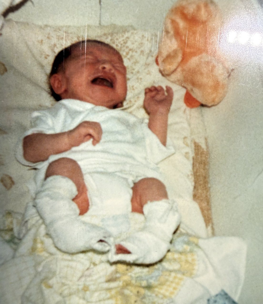
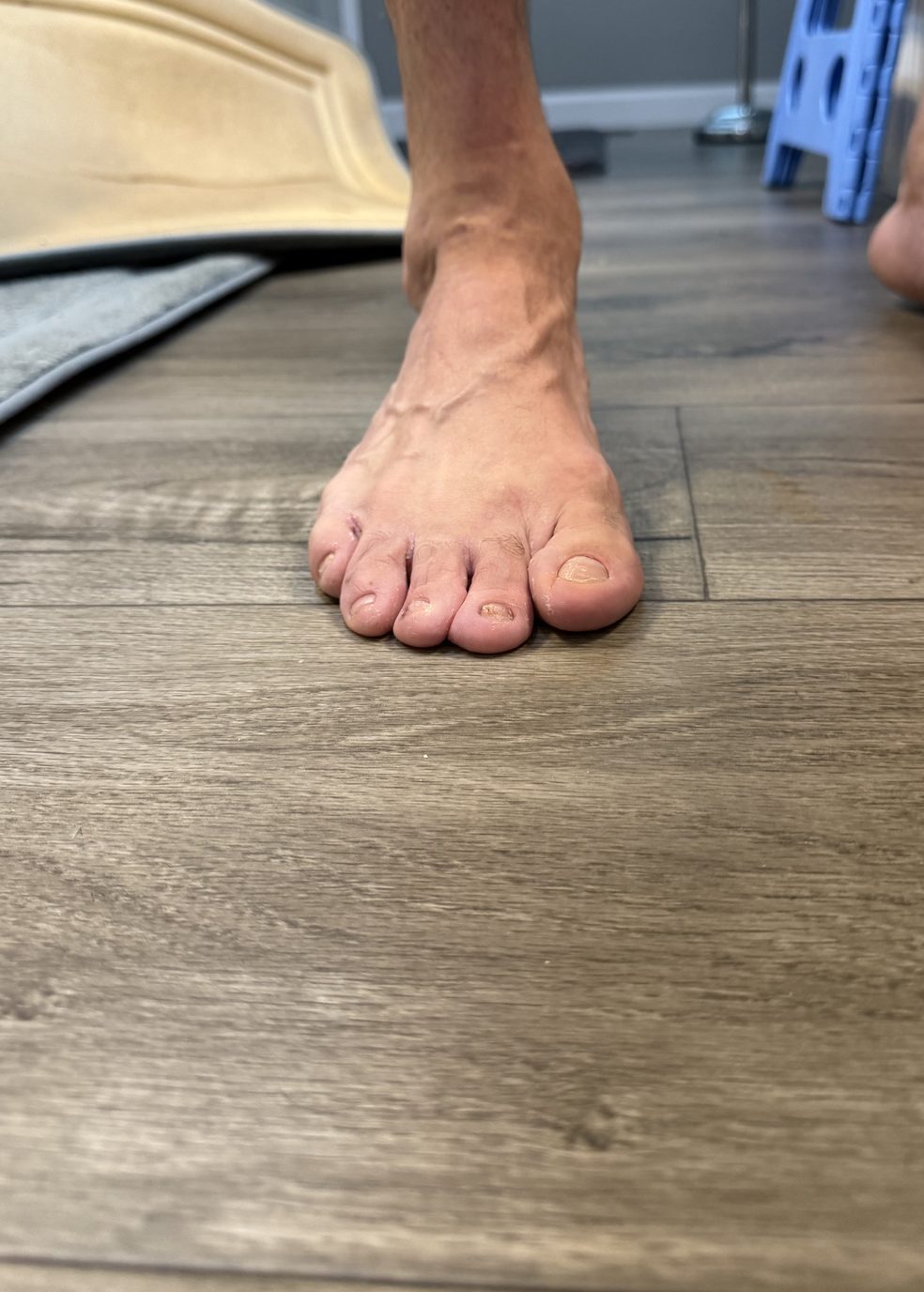
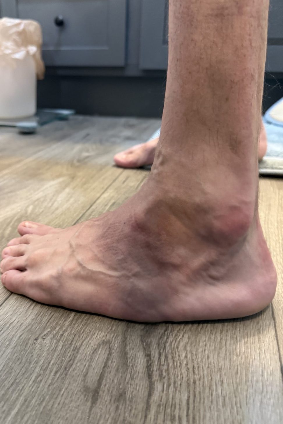
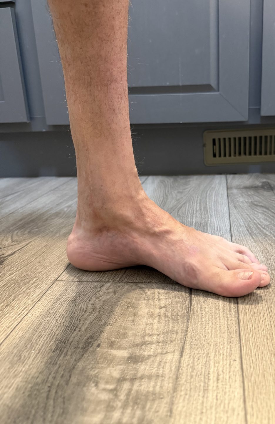
This page is built from a real adult bilateral clubfoot history where both feet were treated aggressively from infancy, yet residual deformity remained across childhood, adolescence, and adulthood. The left foot repeatedly stayed worse than the right, with persistent supination, hindfoot varus, metatarsus adductus, cavovarus/equinovarus recurrence, limited motion, lateral loading, pain, and eventual triple arthrodesis.
The goal here is to explain residual clubfoot deformity the way adults actually experience it: not as a technical leftover on a chart, but as the structural difference that keeps affecting pain, shoes, walking, running, standing work, military service, and long-term function after childhood treatment is supposedly “done.”
Treated can be functional. Treated does not always mean mechanically normal.Plain-Language Summary
If your real question is why the foot still looks, loads, or feels different after treatment, start with the difference between correction and normal anatomy. Residual deformity lives in that gap.
Functional Reality
Residual deformity does not automatically mean the outcome was a failure. It means the foot may still carry part of the original deformity or treatment history while functioning at a higher level than the untreated structure would have allowed.
E-E-A-T Context
Residual deformity explains why someone can be athletic, active, employed, and highly functional while still having a foot that never fully stopped behaving like a clubfoot foot under load. This page keeps that reality grounded in lived adult function, orthopedic language, and clear scope limits rather than oversimplified before-and-after narratives.
Quick answer: Adult residual clubfoot deformity means the foot stayed mechanically different after treatment. It may include persistent varus, cavus, adductus, supination, equinus, limited ankle or subtalar motion, calf asymmetry, lateral border loading, shoe-fit problems, pain, gait compensation, or later degenerative change.
Jump To
What It Means | Deformity Types | How Records Show It | Left vs Right | Pain and Loading | Gait Compensation | Shoes and Orthotics | When Surgery Comes Up | Parent Takeaways | FAQ
Long-Term Adult Paths
If your real question is what childhood treatment and surgery can mean decades later, these pages build the broader cluster:
What Adult Residual Clubfoot Deformity Means in Plain English
Adult residual clubfoot deformity means the foot was treated, but part of the original shape, alignment, motion problem, or load pattern remained. That does not automatically mean treatment failed. In severe clubfoot, treatment may make the foot far more usable while still leaving behind a mechanically different structure.
In plain English, the foot may still act like a clubfoot foot in key ways even after casting, bracing, surgery, or years of function. It may load on the outside. It may stay stiff. It may point inward. It may have a high arch. It may not dorsiflex normally. It may fit shoes poorly. It may look “corrected enough” but still move differently when the body is tired or under stress.
That is why residual deformity matters so much. It explains the gap between being treated and being mechanically normal.
Common Residual Clubfoot Deformity Patterns in Adults
Residual deformity is not one thing. Adults may have one dominant pattern or several overlapping patterns. The exact mix matters because it changes how the foot loads, where pain appears, what shoes work, and whether later treatment becomes necessary.
Residual Varus
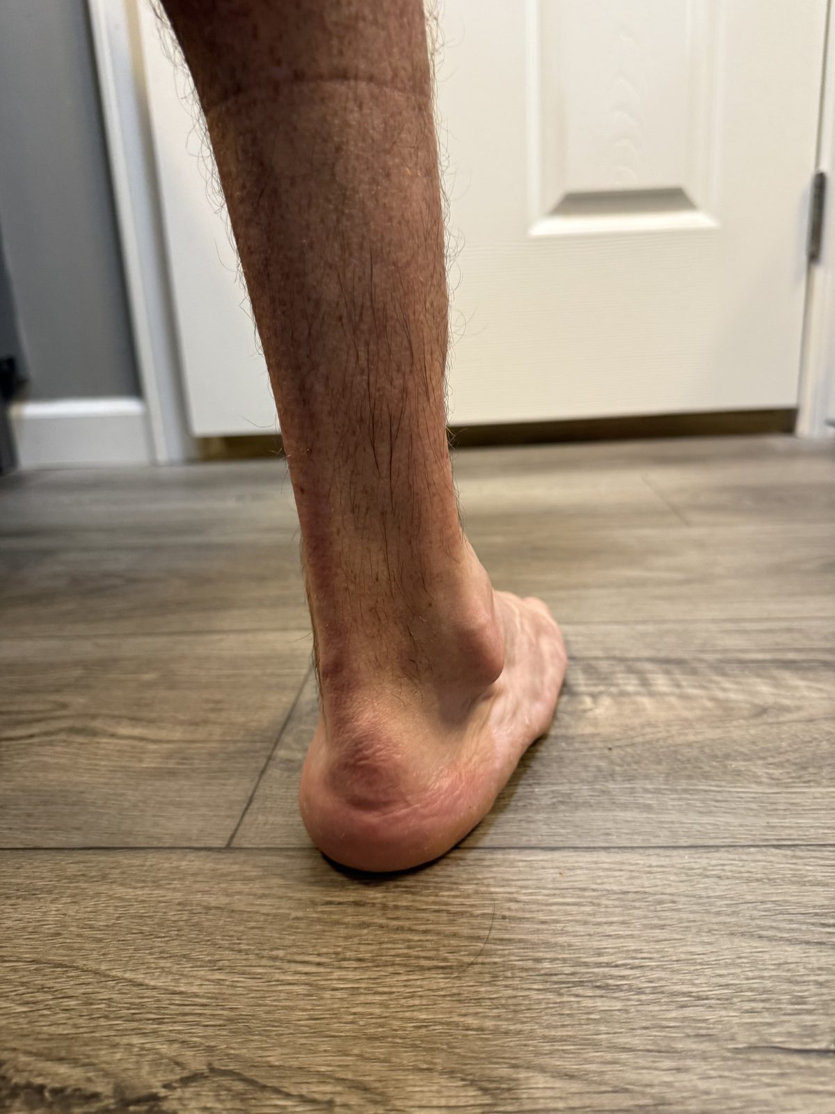
The heel or rearfoot remains tilted inward. This can increase outside-foot loading, outer heel wear, lateral ankle strain, and difficulty keeping the foot flat under load.
Forefoot Adductus
The front of the foot remains pulled inward. This can affect shoe fit, toe-box pressure, walking direction, push-off, and how the forefoot meets the ground.
Cavus or Cavovarus
A high, rigid arch can concentrate pressure through the heel, forefoot, and lateral border. Cavovarus can make the foot feel strong but unforgiving.
Supination Under Load
The foot tends to roll or stay toward the outside edge instead of spreading weight evenly. This can create lateral border pain and stress through the fifth metatarsal region.
Equinus or Limited Dorsiflexion
The ankle cannot move upward normally. This can force early heel rise, forefoot overload, calf fatigue, short stride, stair difficulty, and ankle impingement.
Stiffness and Limited Subtalar Motion
The foot cannot adapt well to uneven surfaces. Missing motion may shift stress into the ankle, knee, hip, lower back, or adjacent joints.
The Important Reframe
Residual deformity does not mean nothing was achieved.
It means treatment improved the foot without erasing the severity of the original problem or the mechanical consequences of the treatment history.
How the Record Actually Shows Adult Residual Clubfoot Deformity
The long-term record does not describe a perfectly corrected foot that later became mysterious. It repeatedly shows residual deformity across years.
Early in the history, the left foot showed supination deformity, hindfoot varus, and metatarsus adductus. The right side functioned better, but it was still not a typical foot. Later notes continued to describe mild heel varus, residual adductus, internal tibial torsion, limited motion, and eventually persistent left cavovarus/equinovarus mechanics.
This is the important long-term point: adult residual clubfoot deformity was not a late surprise. It was a repeated structural theme across the entire timeline. Adult pain and later surgery did not appear out of nowhere. They grew out of a foot that was improved, functional, and still mechanically abnormal.
Why the Left-Worse-Than-Right Pattern Matters
One of the strongest truths in this record is that the left foot kept emerging as the worse side. That matters because adult residual deformity is often not evenly distributed, even in a bilateral case.
The right foot became more plantigrade and functionally better aligned. The left foot kept more varus, more supination, more adductus, more limited motion, more lateral border loading, and more pain with activity. That uneven pattern shaped the entire adult outcome.
Right Foot
More Functional, Still Not Normal
The right foot became more usable and better aligned over time. It still carried clubfoot history, but it did not become the primary breakdown side.
Left Foot
More Deformed, More Costly
The left foot repeatedly stayed worse, more limited, more laterally loaded, and eventually became the side that required triple arthrodesis.
Adult Lesson
Bilateral Does Not Mean Equal
Even when both feet are affected, one side may carry more deformity, more pain, more compensation, and more long-term surgical risk.
How Residual Clubfoot Deformity Turns Into Pain
Residual deformity matters because it changes how the foot loads and moves over years. The issue is not only the shape of the foot. It is what the shape forces the body to do repeatedly.
In this history, the left foot eventually showed significant supination, hindfoot varus, prominent lateral loading, callus formation, recurrent ankle issues, and a fourth metatarsal stress fracture. That is what can happen when a structurally different foot stays athletic and highly functional for years while paying a hidden mechanical cost.
Residual deformity can concentrate stress in predictable areas:
- Heel: uneven rearfoot contact, heel varus, impact intolerance, or plantar fascia strain.
- Ankle: impingement, stiffness, limited dorsiflexion, or arthritic joint loading.
- Arch and midfoot: cavus pressure, rigid midfoot strain, or compensatory collapse around stiffness.
- Outside foot: lateral border loading, fifth metatarsal stress, cuboid pain, or peroneal tendon overwork.
- Forefoot: early heel rise, metatarsal overload, toe-box pressure, or callus formation.
- Calf: overwork from limited ankle motion, weak push-off, or Achilles tightness.
- Knee, hip, and lower back: compensation when the foot and ankle cannot absorb or distribute force normally.
For the location-specific breakdown, continue with Adult Clubfoot Pain by Location.
Residual Deformity and Adult Gait Compensation
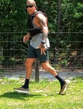
Residual clubfoot deformity often shows up most clearly when the person is tired, walking fast, running, climbing stairs, working on hard floors, or moving across uneven ground. The gait may look “normal enough” during casual walking, but compensation becomes more obvious when demand rises.
Common compensation patterns include:
- shorter stride because the ankle cannot move through full dorsiflexion
- early heel rise because the calf and ankle cannot lengthen normally
- outside-edge loading because the foot stays supinated or varus
- hip rotation or trunk compensation to clear the foot during swing
- reduced push-off because the foot cannot act like a normal lever
- uneven cadence or fatigue-driven limping late in the day
That is why residual deformity is not only a foot topic. It becomes a whole-chain topic. Read the deeper mechanical cluster at Adult Clubfoot Gait Compensation Hub.
Residual Deformity, Shoes, and Orthotics
Residual clubfoot deformity can make ordinary shoe advice fail. A foot with varus, cavus, adductus, stiffness, fusion history, or lateral loading does not always tolerate the same shoe shape, arch profile, heel counter, outsole, or insert that works for a typical foot.
The problem is not only comfort. It is whether the shoe and support system make the loading pattern better or worse. A shoe can feel soft while allowing too much instability. An orthotic can add support while crowding the foot. A narrow toe box can aggravate forefoot adductus. A flexible outsole can feel light while forcing the foot to work harder than it can tolerate.
Adults with residual deformity should pay attention to:
- outside heel and lateral outsole wear
- pressure over bony prominences
- toe-box crowding from forefoot shape
- arch pain from mismatched support
- instability from overly soft shoes
- reduced pain when the shoe adds structure without crushing the foot
For the full footwear strategy, read Adult Clubfoot Shoes and Orthotics and Best Work Shoes for Adult Clubfoot.
When Adult Residual Clubfoot Deformity Raises Surgery Questions
Adult residual clubfoot deformity does not always lead to later surgery. Many adults manage symptoms with shoes, orthotics, bracing, physical therapy, activity modification, load management, and careful monitoring. But residual deformity is one of the clearest reasons later surgery can enter the adult clubfoot conversation.
In this history, the left foot stayed functional for years before the deformity became painful enough, mechanically costly enough, and limiting enough to justify triple arthrodesis. That is the key lesson: residual deformity can be tolerated for a long time before it becomes the central problem.
Surgery discussions become more relevant when:
- pain escalates instead of settling
- lateral border loading becomes a repeat structural pattern
- stress injury or overload signs start appearing
- ankle, hindfoot, or midfoot arthritis becomes function-limiting
- walking, work, or activity tolerance keeps declining
- conservative management stops controlling symptoms
- gait compensation starts creating problems higher up the chain
If that sounds familiar, continue with Revision Clubfoot Surgery in Adulthood, Adult Clubfoot Surgery Later in Life, and Triple Arthrodesis for Clubfoot: Real Long-Term Outcome.
Why Adults Often Discover This Concept Late
Many adults are told as children that their feet were treated, corrected, or handled. They are not always told that residual deformity may still remain in a structural and mechanical sense.
That gap in explanation can leave adults confused years later. They may wonder why one foot still feels different, why shoes are difficult, why running feels expensive, why standing work causes unusual fatigue, or why pain appears in a pattern that standard advice does not explain.
Residual deformity gives language to something many adults have felt for years without being able to name clearly.
What Parents Should Actually Take From This
Parents should not read adult residual clubfoot deformity as automatic failure. The real lesson is more nuanced. A severe foot can be treated, improved, and made highly functional while still remaining structurally different. Both things can be true at the same time.
The useful takeaway is long-term honesty. A child can be active, athletic, and strong for years while still carrying residual deformity that may matter later. That does not mean parents should panic. It means parents should keep watching gait, shoe wear, pain, stiffness, calf size, activity tolerance, recurrence signs, and whether one side begins falling behind the other.
The goal is not fear. The goal is better long-term tracking.
Pain Progression
Why Pain Gets Worse
Continue with Why Adult Clubfoot Pain Gets Worse Over Time and Adult Clubfoot Pain by Location.
Later Surgery
Revision and Fusion Decisions
Go next to Revision Clubfoot Surgery in Adulthood and Adult Clubfoot Surgery Later in Life.
Adult Residual Clubfoot Deformity FAQ
What is adult residual clubfoot deformity?
It means part of the original clubfoot alignment, shape, stiffness, or loading pattern remains after treatment. The foot may be functional and improved while still being mechanically different.
Does residual deformity mean my childhood treatment failed?
Not automatically. Treatment can make a severe clubfoot much more usable without making it normal. Residual deformity may reflect the severity of the original condition, recurrence, growth, treatment limits, surgical history, or long-term stiffness.
What residual deformities can remain after clubfoot treatment?
Residual patterns can include heel varus, forefoot adductus, cavus, supination, equinus, limited ankle motion, limited subtalar motion, calf asymmetry, lateral border loading, internal tibial torsion, and sometimes overcorrection patterns.
Can residual deformity cause adult pain?
Yes. Residual deformity can change how force travels through the foot and leg. Pain may appear in the heel, ankle, arch, outside foot, forefoot, calf, knee, hip, or lower back depending on the compensation pattern.
Can residual deformity affect shoes and orthotics?
Yes. A residual clubfoot shape may not match standard shoe geometry. Some adults need wider toe boxes, deeper shoes, more stable soles, custom orthotics, pressure relief, or careful footwear testing.
When does residual deformity become a surgical issue?
It becomes more concerning when pain is worsening, gait is changing, lateral loading is creating stress injury, arthritis is limiting function, conservative care is failing, or daily life and work tolerance are declining.
Related Pages
- Adult Clubfoot Life Hub
- Long-Term Effects of Childhood Clubfoot Surgery
- Revision Clubfoot Surgery in Adulthood
- Why Adult Clubfoot Pain Gets Worse Over Time
- Adult Clubfoot Operative History and Long-Term Function
- Triple Arthrodesis for Clubfoot: Real Long-Term Outcome
- Adult Clubfoot Work Survival System
External Medical References
These sources provide broader medical context for residual, recurrent, late, and adult clubfoot deformity patterns:
- PubMed: The Adult Sequelae of Treated Congenital Clubfoot
- PMC: Long-Term Follow-Up of Children With a Surgically Treated Clubfoot
- PMC: Correcting Residual Deformity Following Clubfoot Releases
- PMC: Late Effects of Clubfoot Deformity in Adolescent and Young Adult Patients
- PMC: Arthrodesis of the Foot or Ankle in Adult Patients With Congenital Clubfoot
- AAOS OrthoInfo: Clubfoot Clinical Overview
Where to Go Next
If this page helped explain why a treated foot can stay structurally different for years, the next best step is the pain progression page or the revision surgery page depending on whether your main question is symptoms, mechanics, or later treatment.
Continue with Why Adult Clubfoot Pain Gets Worse Over Time or Revision Clubfoot Surgery in Adulthood.
Critical Disclaimer
This page shares educational summaries and lived-experience framing only. It is not medical care, diagnosis, or individualized treatment. Questions about deformity, pain, gait change, function, orthotics, imaging, or later surgery should be discussed with a qualified orthopedic specialist, podiatrist, physical therapist, or medical professional who understands your exact clubfoot history. For site standards, see the Clubfoot Editorial Policy.