
What a treated clubfoot actually looks like, at forty-one
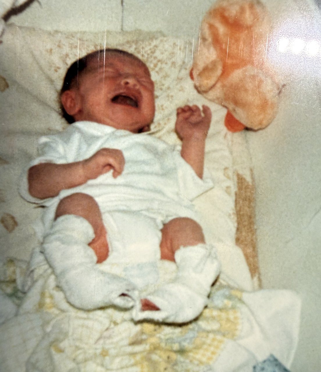
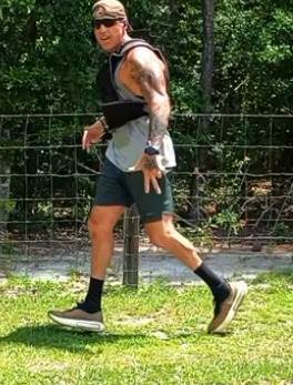
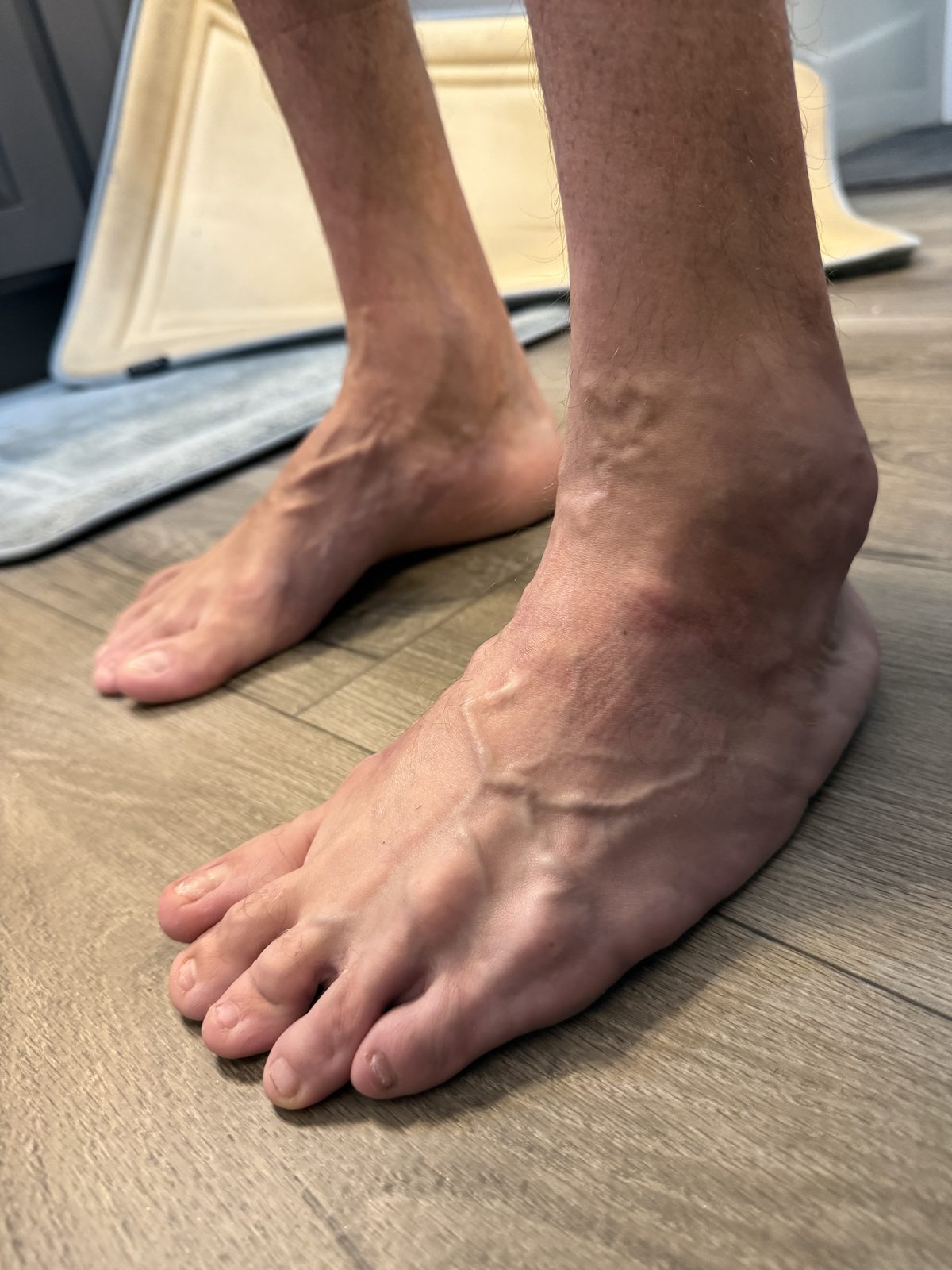
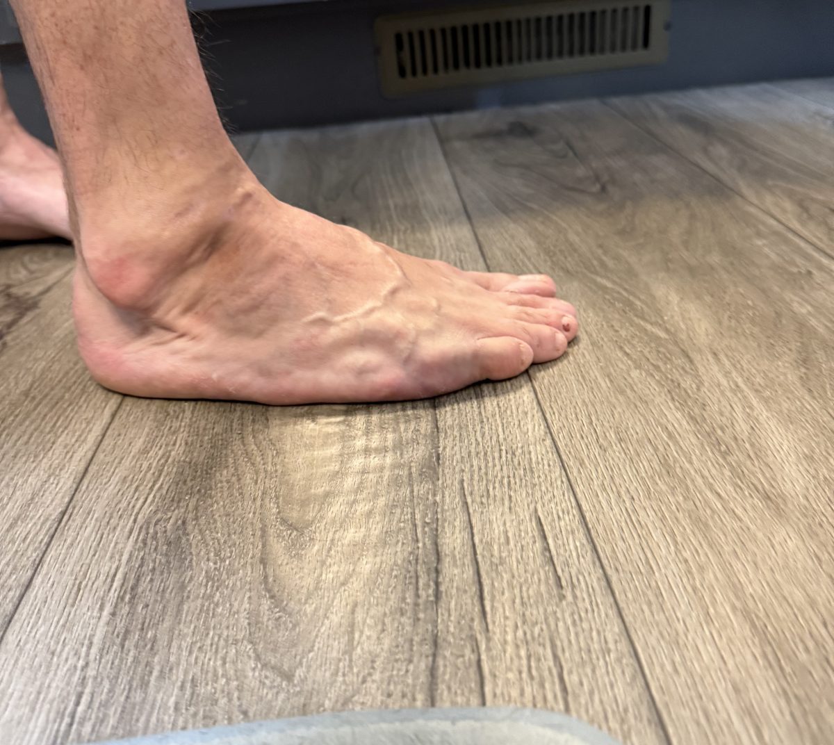
You can find thousands of photographs of an untreated clubfoot on a newborn. You will struggle to find any of a treated one on a middle-aged adult. That absence is why people end up comparing their own feet against a picture of a condition they no longer have. These are mine, bilateral, casted from birth, operated on at four months, fused on the left at sixteen.
Start here
Why you cannot find these pictures
Medical photography follows the condition, not the person. Pictures get taken to document a deformity before it is corrected, and to show the result a few months or a few years after. Then the reason to take them runs out. Nobody photographs a foot at forty-one, because by then there is no clinical question that a photograph answers.
So the image sitting at the top of every search result is a newborn, untreated, feet turned in hard. That image is doing real harm to adults, because it is the thing they measure themselves against, and it is a picture of something their foot has not been since before they can remember.
The honest inventory
What you actually see
Four things, and only the first is unique to having been operated on.
Each of those has its own page with the evidence on it. This page is the part those pages cannot do, which is show you.
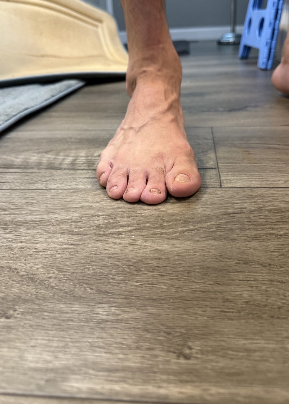
Three scars, three operations
The marks, and what each one is
I could not have told you what any of these were until this month, when I finally read my own operative notes. I had carried them for four decades and known them only as the shape of my own legs.
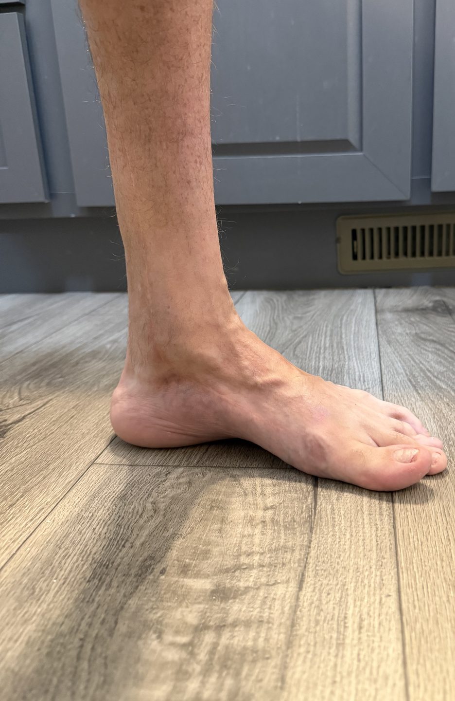
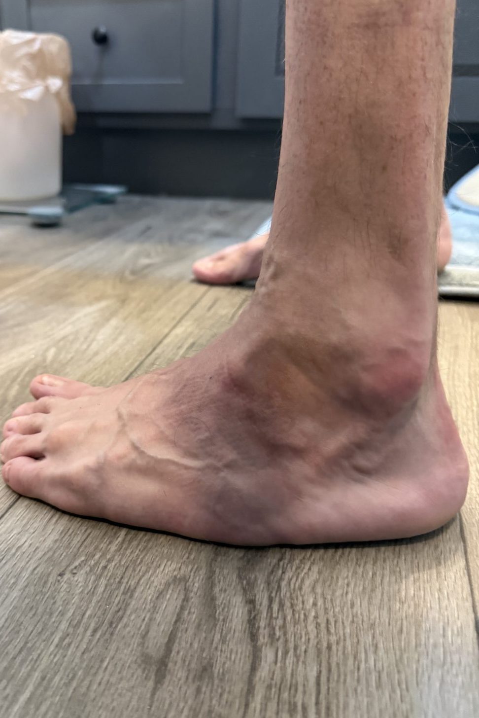
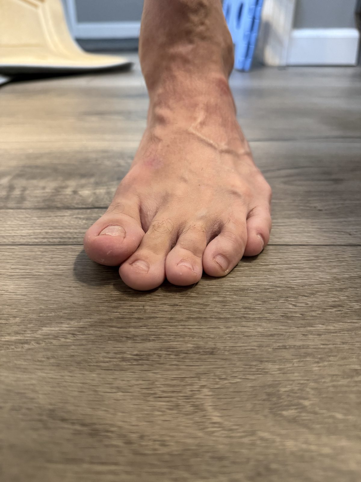
If you were treated before roughly the 1990s, extensive surgical release was the standard approach and your scars will look broadly like mine. If you were treated after, with casting, you may have one small mark at the back of the heel and nothing else. The scars date you more accurately than the foot does.
The limit of a photograph
What none of this can tell you
This is the part that matters most and it is the part a picture is worst at.
If you came here to find out whether your foot looks the way it should, the honest answer is that I cannot tell you, and neither can a photograph of somebody else. What a picture is genuinely good for is the opposite question. Not is mine normal, but am I the only one. You are not.
The gap
The part I cannot photograph
The thing that actually hurts barefoot is not in any of these pictures.
There is a bone on the outer side of my midfoot that takes weight it was not designed to take. I have always assumed it is the base of the fifth metatarsal, standing proud because of the way the foot curves inward, and a clinic note from when I was fifteen does record a prominent and painful spot in the same place. Nobody has ever examined it for that as an adult and no imaging identifies it, so I am not going to state the bone as fact on a page people may take to an appointment.
I also cannot show it to you, which took me a while to work out. The border rolls under, so from the side and from above there is nothing to see. It only appears from underneath, and you cannot hold a camera and lift your own foot at the same time.
People also ask
What adult clubfoot looks like
What does clubfoot look like in adults?
Do adults with clubfoot have scars?
Can you tell someone had clubfoot by looking at their feet?
Does a treated clubfoot look normal?
Why is one clubfoot foot smaller than the other?
Provenance
Where this comes from
The photographs are mine, taken in September 2026, unretouched apart from cropping and resizing. I am forty-one, with bilateral clubfoot recorded as congenital talipes equinovarus, casted from the birth admission, operated on at four months and fused on the left at sixteen.
The dates attached to each scar come from my own operative records, which I hold: the Sarasota admission of July 1985 and the Shriners file covering 1988 and 2001. Which scar belongs to which operation is my identification, matched against what those notes say was done and where.
The general statements on this page about calf size, foot size and residual components are not sourced here. They are covered, with their citations, on the pages linked above, and I would rather send you to the evidence than restate it badly.
Nothing on this page is a clinical reading of a photograph. The only time a clinician has examined or X-rayed either foot in my adult life was an urgent care visit in 2023 for a bruise, which was not about any of this, so any sentence that would require an examination to support it is not here.
Keep going
Related
Written by Heath, founder of Clubfoot Forward, an adult with bilateral clubfoot, extensive childhood surgery, a left-sided relapse and a triple arthrodesis. Photographs of his own feet, published so that adults searching for this have something to look at other than a newborn. Lived experience and orientation, not medical advice, and not a substitute for having your own foot examined. Reviewed September 2026. See the editorial policy.